CT Scanner
MBH-60
DESCRIPTION:
FEATURES
IMPROVED X-RAY EFFICIENCY
- Lower image noise and fewer artifacts
- Reduced signal loss and higher image resolution
- Higher SNR (Signal-to-Noise Ratio) with lower radiation dose
- The neurovascular system, as well as the small vessels of the renal and pulmonary arteries, can be clearly visualized.
SAFE, EFFICIENT, LOW DOSE
- Automatic patient positioning
- Automatic table movement to the scan center
- Improves scanning efficiency
- Remote scanning capability
- Easy to learn and operate
- Reduces radiation dose
- Standardized workflow protocols
LOW-DOSE TECHNOLOGY
- iDream IR (Iterative Reconstruction)
- V-Dose Check
- V-Dose Report
- Low-kV Scanning
- Automatic mA (Auto mA)
- Pediatric Protocol
- V-Beam Collimator Tracking
COMPREHENSIVE APPLICATIONS
Interventional Department: Biopsy, ablation, and treatment procedures.
Radiotherapy Department: CT simulation.
Oncology Department: Tumor imaging, evaluation, follow-up, and treatment assessment.
Orthopedics Department: 3D and VR imaging for fracture visualization.
Medical Examination Center: Low-dose screening and automated diagnosis.
Neurology Department: Stroke evaluation and vascular morphological analysis.
Respiratory Department: Lung nodule analysis, pneumonia detection, and pulmonary function assessment.
Radiology Department: Full-body scanning.
TECHNICAL
SPECIFICATIONS

GANTRY
Bore Size: 75 cm
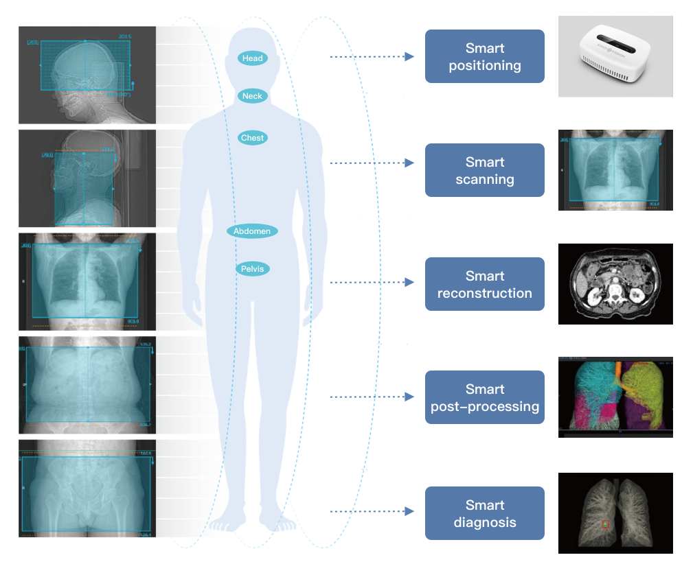
One-Touch Positioning: 3 preset protocols
Scan Speed / 360°: 0.7, 0.8, 0.9, 1.0, 2.0 s
Scan Field of View: 50 cm
Tilt Range: Mechanical tilt ±45° (0.1° step)
Display Panel
Size: 13.3-inch LCD displaying current scan parameters
Automatic Voice Guidance: Supported
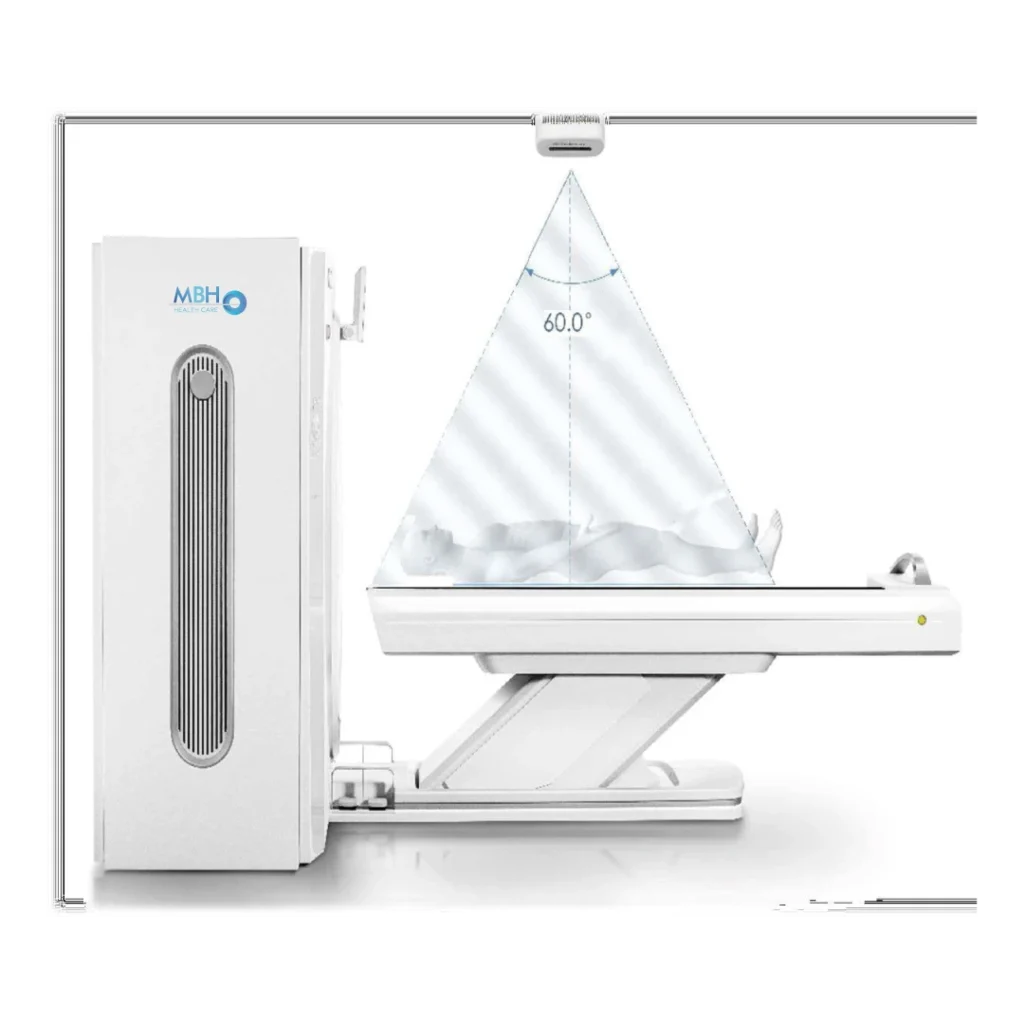
3D Vision Camera: Optional

PATIENT TABLE
Maximum Horizontal Travel Range: 1600 mm
Scannable Horizontal Range:
Scout Scan: 50 – 570 mm
Axial Scan: 1570 mm
Helical Scan: >1550 mm
Horizontal Movement Speed: 1–200 mm/s
Vertical Table Travel Range: 510 mm – 885 mm
Maximum Table Load: 210 kg
One-Touch Patient Table Release: Supported
Patient Table Support Switches: Supported

X-RAY TUBE
Anode Heat Capacity: 3.5 MHU
Equivalent Anode Heat Capacity: 9.5 MHU (with iDream)
Cooling Rate: 735 kHU/min
Focal Spot Size:
Large: 1.2 mm × 1.4 mm
Small: 0.7 mm × 0.8 mm

GENERATOR
Power Output: 42 kW
Equivalent Rated Power: 113 kW (with iDream)
kV Settings: 70, 80, 100, 120, 140 kV
mA Range (Step Size): 10–300 mA (1 mA step)

DETECTORS
Material: Solid-State GOS
Number of Detector Rows: 32 rows
Maximum Slices per Rotation: 64 (conjugate mode)
Detector Channels per Row: 864
Total Detector Elements: 27,648
Minimum Slice Thickness: 0.3125 mm
Detector Width: 20 mm
Maximum Data Sampling Rate: 4800 views / 360°

SCANNING PERFORMANCE
Scout Scan: Supports three modes: AP, lateral, and dual; scannable range 50–1570 mm.
Acquisition Modes
64 × 0.625 mm
32 × 0.625 mm
16 × 0.625 mm
2 × 0.3125 mm
Minimum Slice Thickness: 0.3125 mm
Collimation Width Selection: 20 mm, 10 mm, 0.625 mm
Pitch Factor: 0.2–2.0 (multiple selections)
Maximum Continuous Scan Time: 100 s

IMAGE RECONSTRUCTION
Reconstruction Field of View (FOV): 50–500 mm; 50–650 mm (extended)
Reconstruction Matrix: 512 × 512, 768 × 768, 1024 × 1024
Reconstruction Speed: ≥ 40 fps (images per second)
Display Matrix: 1024 × 1024

IMAGE OPTIMIZATION ALGORITHMS
Metal Artifact Reduction: Standard
Beam Hardening Artifact Reduction: Standard
Partial Volume Artifact Reduction: Standard
Ring Artifact Reduction: Standard
Helical Scan Artifact Reduction: Standard
Motion Artifact Reduction: Standard

IMAGE QUALITY
Spatial Resolution: ≥ 21 lp/cm @ 0% MTF; XY plane: ≥ 15 lp/cm @ 0% MTF; Z plane
Low-Contrast Resolution: 2 mm @ 0.3% @ 25 mGy
Image Noise: < 0.35% (central dose of 40 mGy)
CT Number Scale (HU):
Standard: –1024 HU to +3072 HU
Extended: –32768 HU to +32767 HU
IMAGE RECONSTRUCTION
Reconstruction Field of View (FOV): 50–500 mm; 50–650 mm (extended)
Reconstruction Matrix: 512 × 512, 768 × 768, 1024 × 1024
Reconstruction Speed: ≥ 40 fps (images per second)
Display Matrix: 1024 × 1024
IMAGE OPTIMIZATION ALGORITHMS
Metal Artifact Reduction: Standard
Beam Hardening Artifact Reduction: Standard
Partial Volume Artifact Reduction: Standard
Ring Artifact Reduction: Standard
Helical Scan Artifact Reduction: Standard
Motion Artifact Reduction: Standard
IMAGE QUALITY
Spatial Resolution: ≥ 21 lp/cm @ 0% MTF; XY plane: ≥ 15 lp/cm @ 0% MTF; Z plane
Low-Contrast Resolution: 2 mm @ 0.3% @ 25 mGy
Image Noise: < 0.35% (central dose of 40 mGy)
CT Number Scale (HU):
Standard: –1024 HU to +3072 HU
Extended: –32768 HU to +32767 HU
COMPUTER SYSTEM:
Hard Drive: 4 TB
(System disk: 0.3 TB + image disk: 1.7 TB + raw data disk: 2 TB)
Monitor:
Size: 24-inch LCD
Resolution: 1920 × 1200
Brightness: 600 cd/m²
Contrast Ratio: 1000:1
Image Storage Capacity: ≥ 3,200,000 images (512 × 512)
External Storage: DVD/CD-RW, USB
Print Interface: Standard DICOM 3.0
CPU: Intel Xeon, 4 cores / 8 threads, 3.6 GHz frequency, 8.25 MB cache
RAM: 32 GB ECC
Dedicated Pediatric Protocols: Standard
Auto mA: Standard
V-Dose Check: Standard
Low-Dose Lung Screening: Standard
240° Exposure: Standard
V-Beam Technology: Standard
V-Dose Report: Standard
iDream Iterative Reconstruction: Standard
V-Bolus Tracking: Standard
V-Bolus Timing: Standard
STANDARD ACCESSORIES
Table Pad
Headrest
Headrest Cushion
Lower Front Belt
Lower Front Belt
Lower Front Belt
Chest and Abdomen Belt (Standard)
Chest and Abdomen Belt (Narrow 1)
Chest and Abdomen Belt (Narrow 2)
Water Phantom
System Phantom
Phantom Holder
OPTIONAL ACCESSORIES
Table Extension
Knee Cushion
Head and Arm Support
IV Pole
Flat Table Top
Laser Positioning Light
3D Vision Camera
Wired Motion Control Panel
DIMENSIONS AND WEIGHT
Patient Table
Width: 2400 mm.
Height: 950 mm.
Weight: 350 kg.
Gantry
Width: 940 mm.
Height: 1850 mm.
Weight: 1200 kg.
Console
Width: 630 mm.
Height: 630 mm.
Weight: 50 kg.
Be part of this great family that is always committed to delivering the very best for you.
Our clients are always our top priority.
OPERATING ENVIRONMENT:
Scan Room Dimensions
Minimum Area: 18 m² (5000 mm × 3600 mm)
Recommended Room Size: 30 m² (6000 mm × 5000 mm)
Recommended Height: 2650–3050 mm (if the 3D Vision Camera is selected)
Temperature:
Scan Room: 20–26 °C
Control Room: 18–28 °C
Humidity:
Scan Room: 30%–70%, non-condensing
Control Room: 20%–80%, non-condensing
POWER SUPPLY REQUIREMENTS
Power Supply Option: Three-phase 380 VAC, voltage variation tolerance < ±10%.
Frequency: 50 Hz or 60 Hz, tolerance < ±1 Hz

