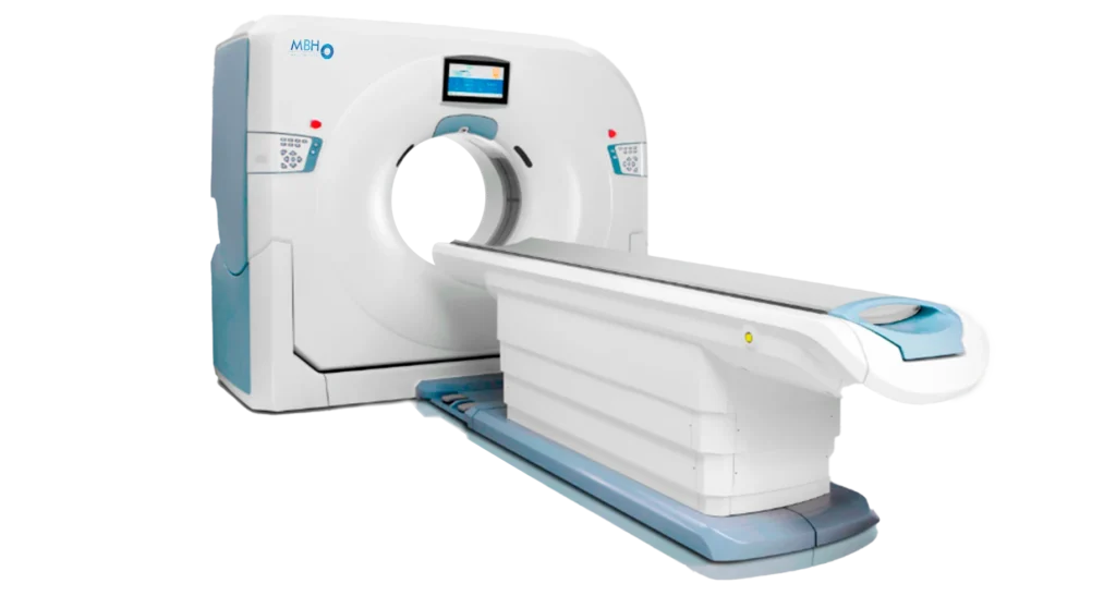
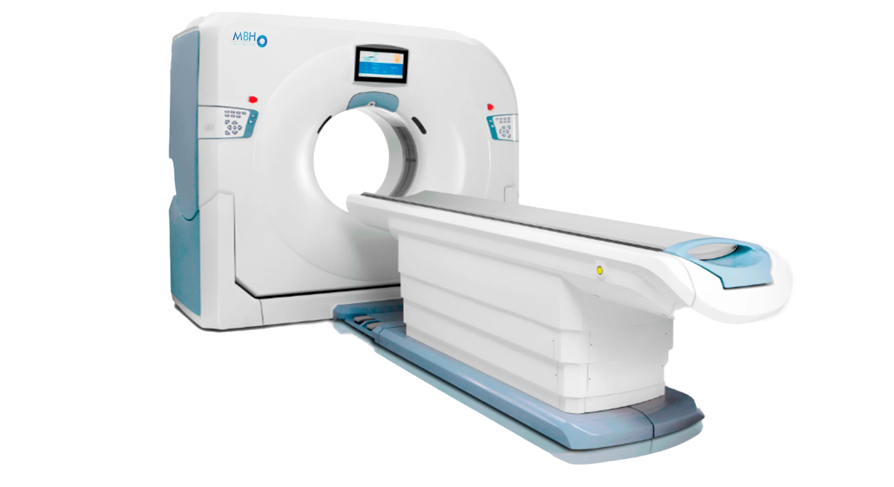
GANTRY
Bore Size: 76 cm
One-Touch Positioning: 3 preset protocols
Scan Speed / 360°: 0.5, 0.6, 0.7, 0.8, 0.9, 1.0, 2.0 seconds
Scan Field of View (FOV): 50 cm
Tilt Range: Mechanical tilt ±30° (0.5° step)
Display Panel: 13.3-inch LCD screen displaying current scan parameters
Automatic Voice Commands: Supported
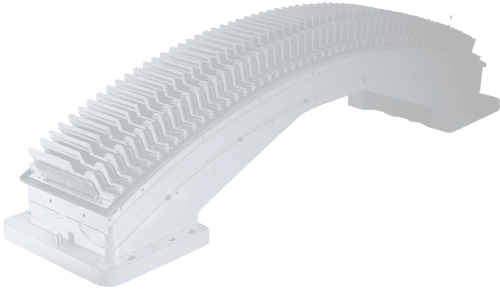
DETECTOR
Material: Solid-State GOS
Number of Detector Rows: 32 rows
Maximum Slices per Rotation: 32
Detector Channels per Row: 864
Total Detector Elements: 27,648
Minimum Slice Thickness: 0.625 mm
Detector Width: 20 mm
Maximum Data Sampling Rate: 4800 views / 360°
PATIENT TABLE
Maximum Horizontal Travel Range: 1950 mm
Scannable Horizontal Range: 50 mm – 1860 mm
Horizontal Movement Speed: 1 mm/s – 200 mm/s
Vertical Table Travel Range: 425 mm – 990 mm
Maximum Table Load: 250 kg
One-Touch Patient Table Release: Supported
Patient Table Base Switches: Supported
GENERATOR
Power Output: 50.4 kW
mA Range: 10–420 mA
kV Settings: 70, 80, 100, 120, 140 kV
X-RAY TUBE
Anode Heat Capacity: 5.3 MHU
Cooling Rate: 815 kHU/min
Focal Spot Size:
Large: 1.0 mm × 1.0 mm
Small: 0.5 mm × 1.0 mm
76 cm
Bore Size
360 °
Scan Speed
250 kg
Maximum Load
5.3 MHU
Heat Capacity
More
Features
DICOM 3.0 interface with bidirectional transmission, compliant with the IHE-C standard, supporting Scheduled Workflow (SWF) and Portable Data for Imaging (PDI) image processing.
REQUIREMENTS
POWER
CONSOLE
MBH-32
• Anzeigematrix: 1024×1024
• Rekonstruktionsgeschwindigkeit: ≥12 fps, tatsächliche Geschwindigkeit bis zu 28 fps
• Monitor: 24,1 Zoll
• Externer Speicher: DVD/CD-RW, USB
• Übertragungsschnittstelle: DICOM 3.0-Schnittstelle, bidirektionale Übertragung, kompatibel mit dem IHE-C-Standard, SWF-Backup-Workflow, portable PDI-Bildverarbeitung
SCANNER
LEISTUNG
• Scanbereich: 50 ~ 1860 mm; 32 x 0,625 mm
• Akquisitionsmodi: 24 x 0,625 mm
• 16 x 0,625 mm
• 8 x 0,625 mm
• Minimale Schichtdicke: 0,625 mm
• Dynamischer Scan: 20-mm-Perfusionsscan
• Wählbare Kollimationsbreite: 20 mm, 15 mm, 10 mm, 5 mm
• Schrittfaktor: 0,2 ~ 1,75 (Mehrfachauswahl)
• Maximale kontinuierliche Scanzeit: 100 Sekunden
We always strive to achieve the best outcome for every patient. Contact us through…
Component Performance
Number of detector rows: 32 rows
Maximum number of slices per rotation: 32
Scan speed / 360°: 0.5, 0.6, 0.7, 0.8, 0.9, 1.0, 2.0 s
Acquisition modes: 8 × 0.625, 16 × 0.625, 24 × 0.625, 32 × 0.625
Tilt range: Mechanical tilt ±30°
Aperture (Bore size): 76 cm
X-ray Tube
Maximum heat capacity: 5.3 MHU
Cooling rate: 815 kHU/min
Environmental Requirements
- Scan Room Temperature: 20–26°C
- can Room Humidity: 30%–70%, non-condensing
- Operating Room Temperature: 18–28°C
- Operating Room Humidity: 20–80%, non-condensing
50 +
hz
S O F T W A R E
» 2D-Bildbetrachter
» 3D SSD
» VR (Volumenrendering)
» Maximum Intensity Projection (MIP)
» Minimum Intensity Projection (MinIP)
» Multiplanare Rekonstruktion (MPR) »
Average Intensity Projection (AIP) »
Virtuelle Endoskopie (VE) »
Auto-mA
iDream Iterative Rekonstruktion »
Spezielle pädiatrische Protokolle
» Bolus-Tracking
» Bolus-Synchronisation V
» Dosisverifizierung V
» Niedrigdosis-Lungenerkennung
» 240 Aufnahmen
V-Beam
V-Dosis-Bericht
Filmdesign und -druck
» Fernkonsultation
Berichtsbearbeitung
Erweiterte Berichtsfunktion
Knochenextraktion mit einem Klick
Energiebedarf
• Leistung 70 kVA
• Dreiphasenspannung 380 V AC, Spannungsabweichung ≤ ±10 %
• Frequenz 50 Hz oder 60 Hz, Toleranz ≤ ±1 Hz
Umweltanforderungen
• Temperatur im Untersuchungsraum: 20–26 °C
• Luftfeuchtigkeit im Untersuchungsraum: 30–70 %, nicht kondensierend
• Temperatur im Operationssaal: 18–28 °C
• Luftfeuchtigkeit im Operationssaal: 20–80 %, nicht kondensierend
Systemleistung
• Scanmodus: Explorer-Scan, Axial-Scan und Spiral-Scan
• Scangeschwindigkeit/360°: 0,5, 0,6, 0,7, 0,8, 0,9, 1,0, 2,0 s
• Abstimmungsfaktor: 0,2–1,75
• Ortsauflösung: ≥ 20 lp/cm, 0 % MTF
Bildoptimierungsalgorithmus
• Reduzierung von Strahlhärtungsartefakten: Standard
• Reduzierung von Partialvolumenartefakten: Standard
• Reduzierung von Streifenartefakten: Standard
• Reduzierung von Spiralabtastartefakten: Standard
• Reduzierung von Bewegungsartefakten: Standard
• Automatische Sprach- und bidirektionale Kommunikation: Integriert in Gantry und Scan-Steuerbox
• Druckschnittstelle: DICOM 3.0n-Standard
Bildqualität
» 20 lp/cm bei 0 % MTF; XY-Ebene
» 15 lp/cm bei 0 % MTF; Z-Ebene
• Niedrigkontrastauflösung: 2 mm bei 0,3 % bei 23,5 mGy;
• Bildrauschen: ≤ 0,35 % (Zentrale Dosis ≤ 26 mGy)
• CT-HU-Skala
• Standard: -1024 HU ~ +3072 HU
• Vergrößert: -32768 HU ~ +32767 HU
Anwendungsgebiete der CTA (Computertomographie-Angiographie)
Anwendungen in der Onkologie
• Kolonanalyse **
• Operationsplanung **
• Tumoranalyse **
Bildrekonstruktion
» 50–500 mm
» 50–650 mm (vergrößert)
• Rekonstruktionsmatrizen: 512x512, 768x768, 1024x1024
• Rekonstruktionsgeschwindigkeit: 12 Bilder pro Sekunde, tatsächliche Geschwindigkeit bis zu 28 Bilder pro Sekunde möglich
• Anzeigematrix: 1024x1024
Anwendungen in der Lunge
• Lungenrundherdkontrastuntersuchung **
• Lungenfunktionsanalyse **
• Erweiterte Lungenrundherdanalyse **
• Erweiterte Pneumonieanalyse **
Neurologische Anwendungen
• RAM: 32 GB DDR4 ECC
• Festplatte: 5 TB (0,3 TB Systemlaufwerk + 0,7 TB Bildlaufwerk + 4 TB Rohdatenlaufwerk)
• Größe: 24 Zoll
• LCD:
» Auflösung: 1920 x 1200
» Helligkeit: 600 cd/m²
» Kontrast: 1000:1
• Bildspeicher: 1.300.000 Bilder (512 x 512)
• Externe Speichermedien: DVD/CD-RW, USB
• Druckerschnittstelle: DICOM 3.0-Standard
Aplicaciones de Neurología
• Digitale Subtraktionsanalyse der Computertomographie **
• Analyse von Schlaganfällen **
Bildoptimierungsalgorithmus
• Reduzierung von Strahlhärtungsartefakten: Standard
• Reduzierung von Partialvolumenartefakten: Standard
• Reduzierung von Streifenartefakten: Standard
• Reduzierung von Artefakten bei Spiralabtastung: Standard
• Reduzierung von Bewegungsartefakten: Standard
• Auflösung bei niedrigem Kontrast: 2,0 mm ± 0,3 %
Dosisoptimierung
• Auto-mA: Standard
• V-Dosis-Verifizierung: Standard
• Niedrigdosis-Lungenscreening: Standard
• 240°-Bestrahlung: Standard
• V-Beam: Standard
• V-Dosis-Bericht: Standard
• Iterative iDream-Rekonstruktion: Standard
• V-Bolus-Tracking: Standard
• V-Bolus-Synchronisation: Standard
Routineanwendungen
• Patienteninformationsmanagement
• DICOM 3.0-Standard
• RIS-Datenmodifikation (Strahleninformationssystem)
• Systemkonfiguration
• Fernkonsultation
• Filmgestaltung und -druck
• Berichtsbearbeitung
• Erweiterte Berichtsfunktionen
• Knochenentfernung mit einem Klick
• 2D-Bildbetrachter
• MPR (Multi-Plane Image Reconstruction)
• 3D, MIP, AIP, MinIP, SSD, VR (Dreidimensionale Bildgebung und Rekonstruktion)
• Patiententischentfernung mit einem Klick
• Virtuelle Endoskopie
Others
Dental analysis, impression modeling, bone density measurement, fat analysis, dual-energy analysis, and rib analysis.

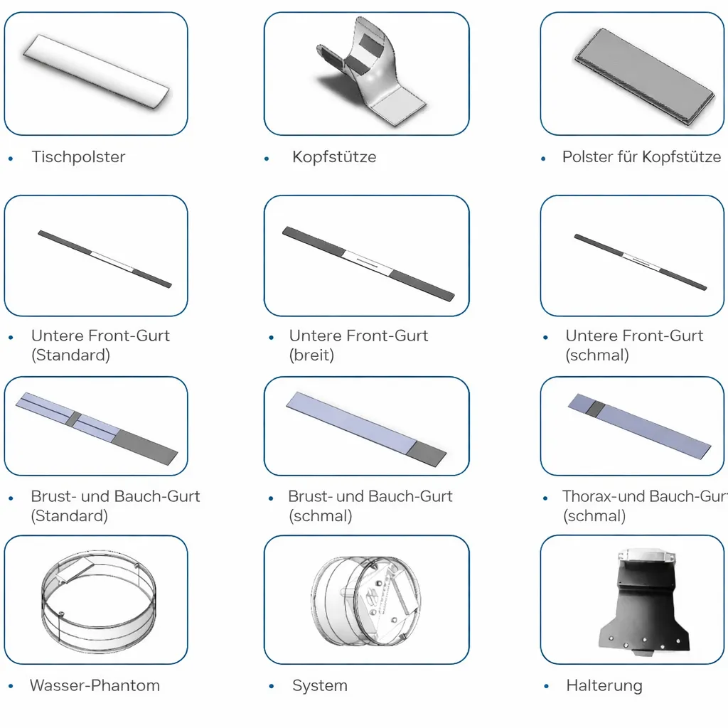
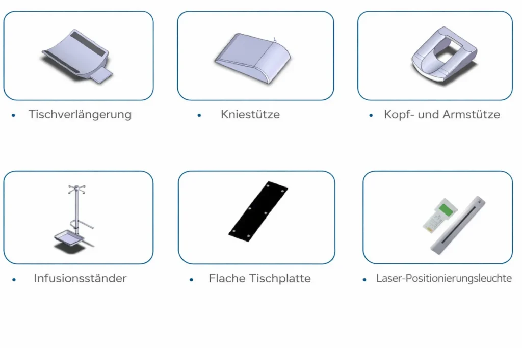
Additional
Zubehör
Operating environment and site requirements, including height and dimensional requirements.
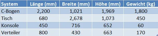
Site Requirements (Recommended)
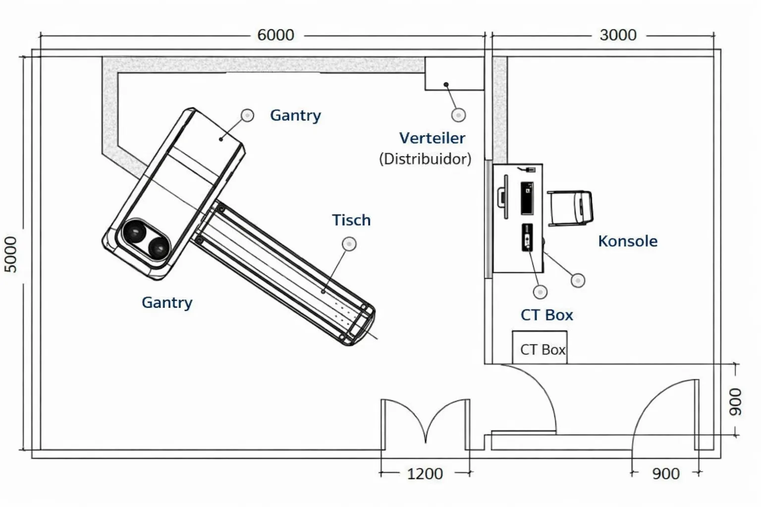
AUS DEM SCANRAUM
ABMESSUNGEN
• Empfohlene Raumgröße: 30 Quadratmeter (6000 mm x 5000 mm)
STROMVERSORGUNG:
ANFORDERUNGEN für
• Stromversorgung: 3-phasig 380 V AC, Spannungsabweichung: Toleranz <±10 %
• Frequenz: 50 Hz oder 60 Hz, Toleranz +1 Hz
UND LUFTFEUCHTIGKEIT:
TEMPERATUR
• Luftfeuchtigkeit: Untersuchungsraum: 30–70 %, nicht kondensierend;
• Operationssaal: 20–80 %, nicht kondensierend
AUS DEM OPERATIONSSAAL
ABMESSUNGEN
INTELLIGENTE ENERGIE:
ERSPARNISSE