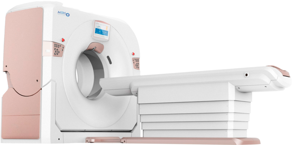
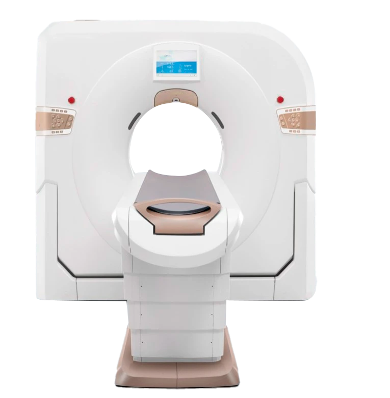
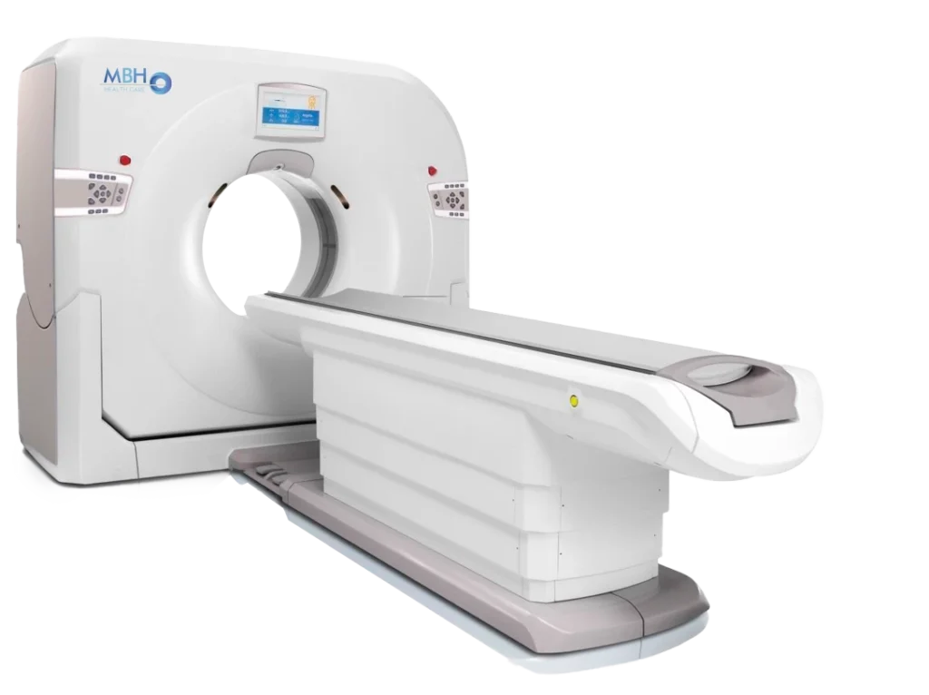
DESCRIPTION
Provides customers with a 128-slice CT scanner featuring a large 76 cm bore, improving image resolution to 21 Lp/cm, enabling more precise disease diagnosis and treatment while significantly enhancing the system’s clinical performance and scalability.
Comprehensive clinical diagnostic platform.
The fully featured 128-slice CT scanner enables accurate localization and diagnosis of general lesions both qualitatively and quantitatively, delivering full clinical value, including extensive cardiac applications supported by an ultra-low dose green platform.
X-RAY TUBE
Anode heat capacity: 5.3 MHU
Cooling rate: 815 kHU/min
Focal spot size:
Large: 1.0 mm × 1.0 mm
Small: 0.5 mm × 1.0 mm
PATIENT TABLE
Maximum travel range (horizontal): 1950 mm
Scannable horizontal range: 50 mm – 1860 mm
Horizontal movement speed: 1–200 mm/s
Vertical table travel range: 425 mm – 990 mm
Maximum table load: 250 kg
One-touch patient table release: Supported
Patient table bracket switches: Supported
GANTRY
Bore size: 76 cm
One-button positioning: 3 preset protocols
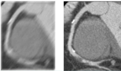
Scan speed / 360°: 0.48, 0.5, 0.6, 0.7, 0.8, 0.9, 1.0, 2.0 s
Scan field of view: 50 cm
Tilt range: Mechanical tilt ±30° (0.5° step)
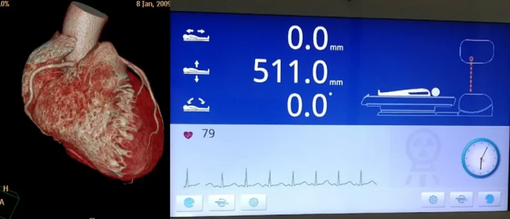
Display panel: 13.3-inch LCD showing current scan parameters
Automatic voice guidance: Supported
ECG cable connection interface: Integrated on the front of the gantry control panel
3D Vision Camera: Optional
GENERATOR
Power output: 50.4 kW
kV settings: 70, 80, 100, 120, 140 kV
mA range (step size): 10–420 mA (1 mA step)
DETECTORS
Material: Solid-state GOS
Number of detector rows: 64 rows
Maximum slices per rotation: 128
Detector channels per row: 864
Total number of detector elements: 55,296
Minimum slice thickness: 0.625 mm
Detector width: 40 mm
Maximum data sampling rate: 4800 views / 360°
SCANNING PERFORMANCE
Scout Scan: Supports three modes: AP, lateral, and dual. Scannable range 50–1860 mm.
Acquisition Modes:
128 × 0.625 mm
64 × 0.625 mm
32 × 0.625 mm
Minimum Slice Thickness: 0.625 mm
Collimation Width Selection: 40 mm, 20 mm, 15 mm, 10 mm
Pitch Factor: 0.2–1.75 (multiple selections)
IMAGE RECONSTRUCTION
Reconstruction Field of View (FOV): 50–500 mm; 50–650 mm (extended)
Reconstruction Matrix: 512 × 512, 768 × 768, 1024 × 1024
Reconstruction Speed: ≥ 60 fps (images per second)
Display Matrix: 1024 × 1024
IMAGE OPTIMIZATION ALGORITHMS
Metal Artifact Reduction: Standard
Beam Hardening Artifact Reduction: Standard
Partial Volume Artifact Reduction: Standard
Ring Artifact Reduction: Standard
Helical Scan Artifact Reduction: Standard
Motion Artifact Reduction: Standard
COMPUTER SYSTEM
CPU: Intel Xeon, 6 cores / 12 threads,
3.8 GHz frequency, 8.25 MB cacheRAM: 32 GB ECC
Hard Drive: 7 TB
System disk: 0.3 TB
Image disk: 1.7 TB
Raw data disk: 5 TB
Monitor:
Size: 24 inches
LCD Resolution: 1920 × 1200
Brightness: 600 cd/m²
Contrast Ratio: 1000:1
Image Storage Capacity: ≥ 3,200,000 images (512 × 512)
External Storage: DVD/CD-RW, USB
Print Interface: Standard DICOM 3.0
CARDIAC SCANNING
Prospective ECG Scanning: Supported
Retrospective ECG Scanning: Supported
Multi-sector Coronary Artery Scanning:
Adaptive sector technology with support for up to 4 sectorsTemporal Resolution:
60 ms; 25 ms @ RTFECG Wave Editing:
Provides editing functions to add or remove abnormal ECG signals (such as premature beats) used for coronary artery recognition.Cardiac Reconstruction:
Automatically selects the optimal reconstruction phase and supports multi-phase reconstruction for a single image.
ADVANCED CARDIAC TECHNOLOGY
RTF Coronary Artery Instant Freeze Technology
(RTF – Real Time Focus)Third-generation motion artifact correction algorithm based on deep learning, capable of modeling and compensating for cardiac motion artifacts. It partially eliminates artifacts caused by heart motion, respiration, and temporal resolution limitations.
Iterative motion displacement correction, repeatedly correcting motion through an iterative process.
Over-correction prevention method, ensuring accurate and stable image reconstruction.
DOSE OPTIMIZATION
Dedicated Pediatric Protocols: Standard
Auto mA: Standard
V-Dose Check: Standard
Low-Dose Lung Screening: Standard
240° Exposure: Standard
V-Beam Technology: Standard
V-Dose Report: Standard
iDream Iterative Reconstruction: Standard
V-Bolus Tracking: Standard
V-Bolus Timing: Standard
ECG mA Modulation: Standard
128-SLICE CT: DESIGNED FOR CARDIAC IMAGING
Fast cardiac scanning in 0.25 seconds
Integrated ECG in the gantry
46 ms cardiac temporal resolution
Prospective or retrospective ECG gating with support for up to four sectors
Large Bore for Expanded Applications
76 cm scanning space, 250 kg table capacity, and 21 Lp/cm resolution make it easy to comfortably accommodate obese patients without compromising radiation dose or image quality.

